The BSF operates several microscopes and image processing computers.
Before using the microscopes, you must sign up here to receive in-person training and to be added to the booking calendar (Exception: Light Microscope and Dissecting Stereoscope).
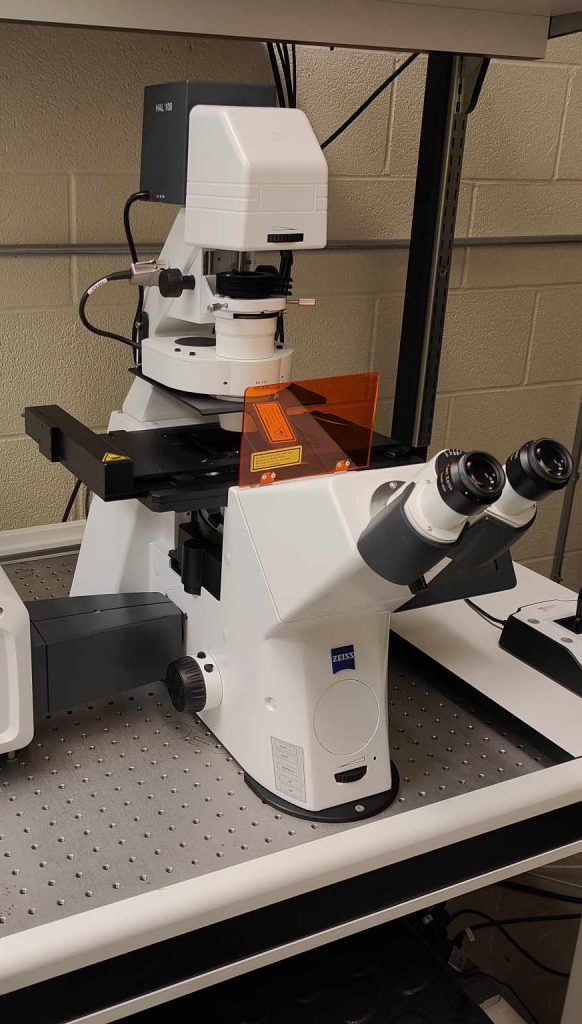
Zeiss Laser Scanning Microscope Confocal Microscope
- Captures multiple high-resolution 2-D images at various depths to reconstruct 3-D structures
- Provides a means of rejecting out-of-focus light from the detector to reduce blur
- Capable of fluorescence and bright-field microscopy
- Equipped with automated cell counter
- Equipped with filters for confocal imagery:
- DAPI (405 nm)
- GFP (488 nm)
- TxRed (594 nm)
- cy5 (635nm)
- Equipped with filters for fluorescence imagery:
- DAPI (405 nm)
- GFP (488 nm)
- Cy3 (555 nm)
- cy5 (635nm)
- Objective lenses:
- 10x, 20x, 40x, 40x (Oil Immersion), 100x (Oil Immersion)
Zeiss Upright Fluorescent/Bright-Field Microscope
- Illuminates specimens with various pre-set wavelengths of light (excitation light) to visualize fluorescence emissions of fluorescent molecules or dyes in the sample
- Capable of slide scanning and image tiling
- Equipped with automated cell counter
- Equipped with a variety of filters:
- DAPI (405 nm)
- GFP (488 nm)
- Cy3 (555 nm)
- TxRed (594 nm)
- cy5 (635nm)
- Objective Lenses:
- 5x, 10x, 20x, 40x (Oil Immersion), 63x (Oil Immersion), 100x (Oil Immersion)
Upright Light Microscopes
- Employs a light source to illuminate samples
- Equipped with a variety of lenses to provide imagery at several magnifications
- Among the most basic of microscopes
- Connected to a computer via HDMI for digital visualization and image-taking of samples
- Objective Lenses:
- 5x, 10x, 20x, 40x, 63x (Oil Immersion)
- Sign-Out Available

Laxco Z200 Dissecting Stereomicroscopes
- Portable dissecting microscopes
- Trinocular viewing system allows for both viewing through lenses and through digital displays
- Standard working distance of 100mm
- 6.4:1 Zoom Ration with 7-45x magnification
- Among the most basic of microscopes
- Sign-Out Available
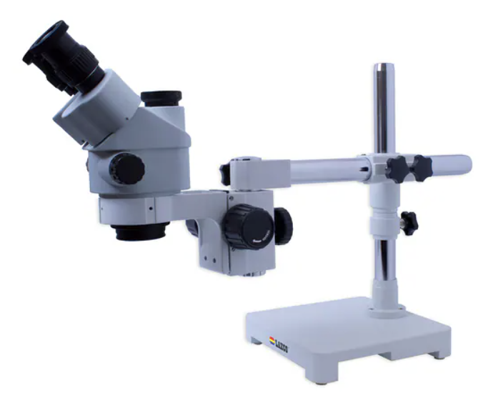
Image Analysis Computers
- Equipped with Zen 2.6 (Blue Edition)
- Can be used in-person or remotely through TeamViewer or Chrome Remote Desktop
CSB Imaging Facility Microscopes
Located in the basement of the Ramsey Wright building, the Cell and Systems Biology (CSB) Imaging Facility operate a variety of microscopes:
- Scanning Electron Microscope (SEM)
- Transmission Electron Microscope (TEM)
- Spinning Disk Confocal Microscope (SDCM)
- Laser Scanning Confocal Microscope (Leica)
Please contact csb.reception@utoronto.ca to learn more about these microscopes.